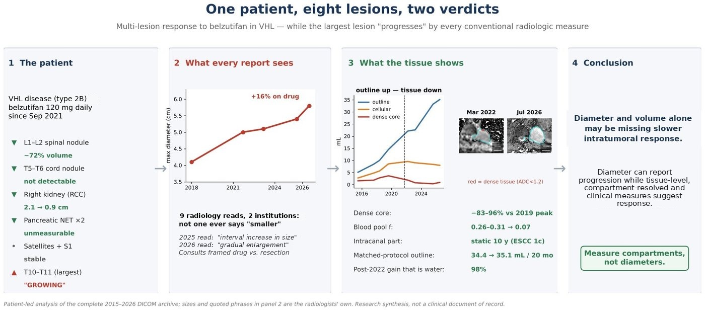
Five lesions responded in ways no one disputes, and two millimeter-scale sentinels never changed. The eighth — the largest — was called “growing” on every conventional read for five years, while the tissue inside it may tell a different story.
Patient TR is an adult male with von Hippel–Lindau disease (type 2B) and roughly three decades of multisystem disease. He has taken the HIF-2α inhibitor belzutifan, 120 mg daily, since September 2021. His complete 2015–2026 imaging archive was re-measured voxel by voxel. RECIST — the Response Evaluation Criteria in Solid Tumors — and the size-based criteria like it were built to judge cytotoxic chemotherapy in malignant tumors: shrinkage is response, growth is failure. This case shows how that ruler may come up short in hemangioblastoma — a contradiction between what the radiology record says about his largest lesion and what the lesion’s interior is doing, on a drug whose earliest effects are vascular rather than cell-killing.
A ruler can only report that this lesion grew, then stopped growing. Compartment-level measurement shows why: diameter and volume alone may be missing slower intratumoral response — the kind of response HIF-2α inhibition was designed to produce.
Within six months of starting belzutifan, each of the five larger non-index lesions in Patient TR’s body lost more than half its volume; the millimeter-scale satellites and the right S1 root lesion, below reliable volumetry, were unchanged. Systemic drug effect is not in question: his hemoglobin has sat at the low plateau that erythropoietin suppression produces for five years (9.9 g/dL, August 2025), and the responding lesions’ regression is radiologist-documented at both treating institutions.
Pre-treatment history relevant here: prior somatostatin-analog therapy 2019–2021 and long-standing off-label propranolol, with quantitatively limited effect on the index lesion.[39] The 1 September 2021 abdominal MRI is the true pre-treatment baseline; the 14 September 2021 spine MRI is approximately day 5 on drug. The lesion-by-lesion record is below. Sizes and quoted phrases in the table are the radiologists’ own; volume percentages are from this analysis.
| Lesion | Pre-treatment (2015–2021) | On belzutifan | Mid-2026 |
|---|---|---|---|
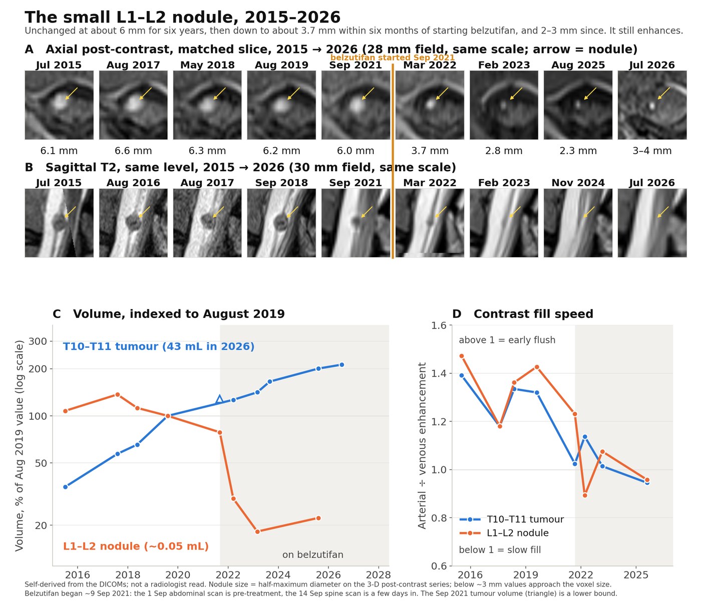
| L1–L2 intradural nodule conus tip, among cauda equina | 6.0–6.6 mm, no growth | 3.7 mm at 6 mo (38% of baseline volume); 23% at 17 mo; 28% at 4 y | 2–4 mm dot, still enhances; best in-CNS sentinel |
| T5–T6 cord-surface nodule + cord edema | ~3 × 5 mm, no growth; edema 6–17 mm | Edema absent from the day-5 scan onward (scanner-era caveat); nodule ~halved by Mar 2022; feeder vessel gone by Aug 2023 | No measurable nodule — cleanest complete response |
| Right kidney mass ccRCC per reports; prior cryoablation ×2, partial nephrectomy | 2.1 × 1.8 cm, “unchanged” vs 2018–2019 (report, Sep 2021) | “Decreased in size from prior,” 1.6 × 1.2 cm at 6 mo (report, Mar 2022); −78% self-derived volume at 17 mo, flat after | 0.9 cm, “no diffusion restricted nodularity” (report, Aug 2025); no identifiable enhancing tissue |
| Pancreatic NET, upper head | 1.1 × 0.8 cm, “unchanged” vs 2018 (report, Sep 2021) | “Smaller and less conspicuous,” 0.7 × 0.6 cm at 6 mo; “no longer discretely measurable” by Feb 2023 | “Not distinctly seen” (report, Aug 2025) |
| Pancreatic NET, uncinate | 12–16 mm (2018–19); 7 mm by Sep 2021 on somatostatin analog | Not measurable on treatment | Not judgeable |
| Millimeter-scale spinal satellites; right S1 root lesion | Unchanged | Unchanged and enhancing | Tracked as sentinels |
| T10–T11 index lesion left extradural paravertebral dumbbell, solid, never cystic | Growing, doubling ~2.8 y; 24.5 mL at drug start (first full-coverage MRI) | Every clinical read reported growth; tissue metrics regressed (section 3) | ~35 mL protocol-locked / ~43–44 mL hi-res full coverage; pain improved; cord unchanged (ESCC 1c stable) |
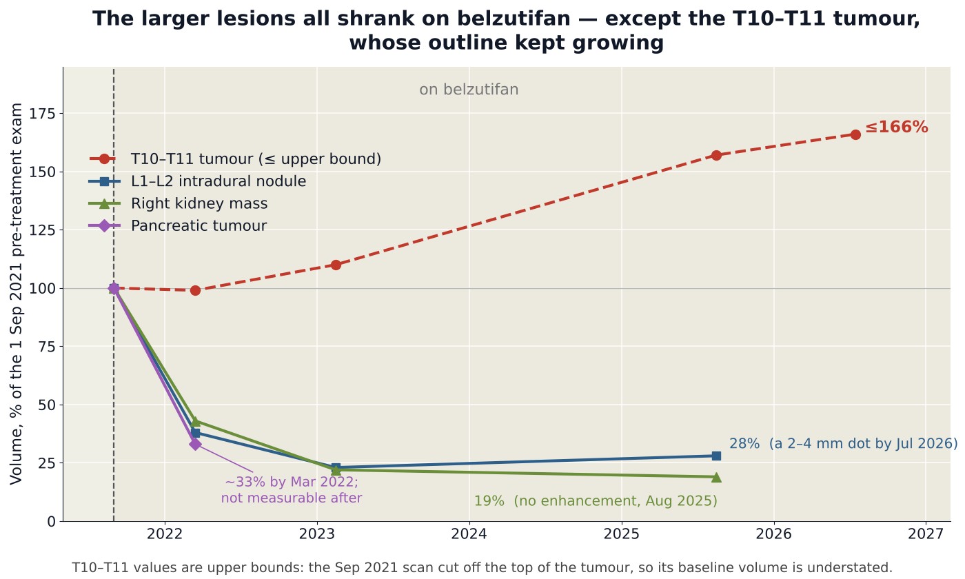
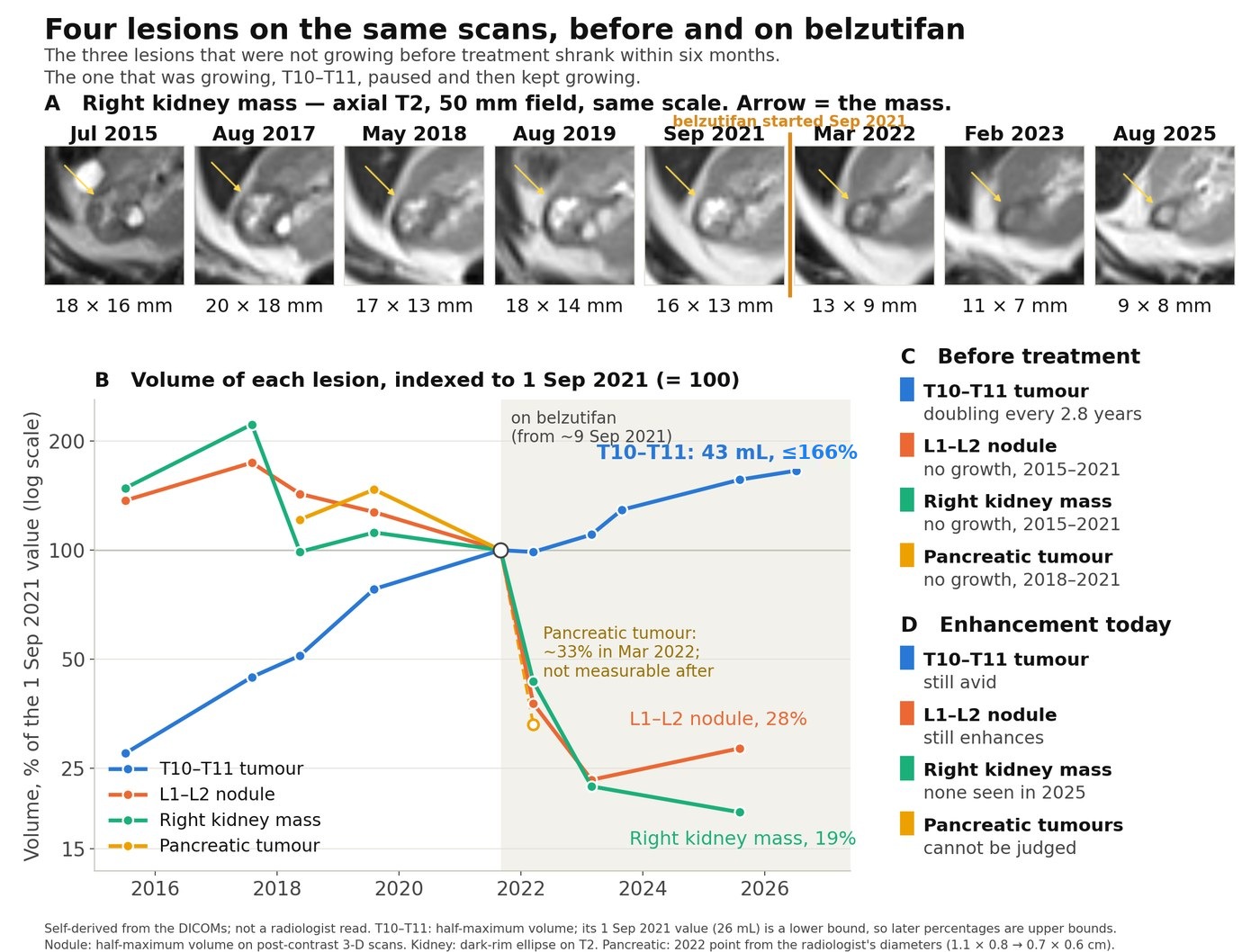
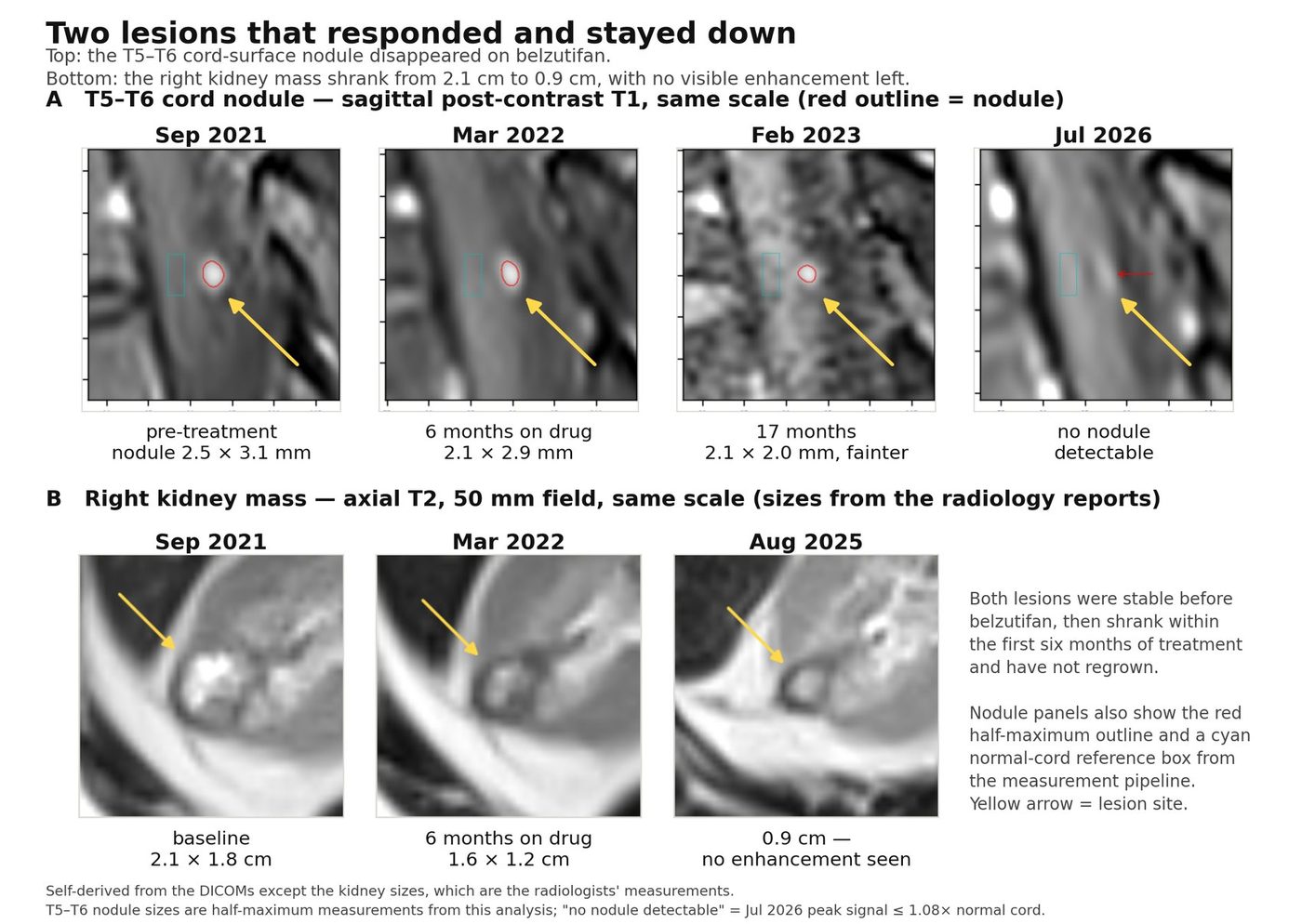
The index lesion is a left T10–T11 extradural “dumbbell” hemangioblastoma at the nerve root — epidural and paravertebral components through the left neural foramen, abutting but not compressing the cord (ESCC 1c — epidural spinal cord compression grade 1c, dural contact without cord deformity — on every exam 2016–2026), solid throughout, with no associated cyst or peritumoral edema on any exam from 2015 to 2026. The institutional record for it, verbatim, is below.
| Exam date | Reported T10–T11 size (radiologist) | Impression wording (verbatim) |
|---|---|---|
| 2018 (reference) | 4.1 × 2.7 × 1.6 cm (spine read); 3.7 × 2.5 cm lateral (abdominal read) | — |
| Sep 1, 2021 | 4.9 × 3.3 cm (lateral) | “…has mildly increased in size” |
| Sep 14, 2021 (day ~5) | 5.0 × 3.2 × 2.9 cm (addendum) | “…appears increase since 2018… Mass effect at the T10 canal appears slightly increased” |
| Mar 16, 2022 | 4.8 × 3.2 × 3.1 cm (spine); 4.5 × 3.4 cm lateral (abdomen) | “…essentially stable in size”; other hemangioblastomas “stable or slightly decreased”; renal mass “decreased in size from prior” |
| Feb 28, 2023 | 5.1 × 3.7 × 3.4 cm | “Mild interval enlargement… Stable mild mass effect upon the T10-T11 cord” |
| Aug 29, 2023 | 5.2 × 3.7 × 3.4 cm | “…stable in appearance” |
| Apr 16, 2024 | 4.0 × 4.9 cm | “Stable exam” |
| Nov 7, 2024 | — (“stable… unchanged”) | “Stable exam since April 16, 2024” |
| Aug 6, 2025 | 5.4 × 4.0 cm | “Interval increase in size of left thoracic paraspinal mass” |
| Jul 10, 2026 | 5.8 × 4.6 cm | “Gradual enlargement of the enhancing mass… Unchanged mass effect on the thecal sac without progressive cord compression or cord edema” |
The stakes were not hypothetical. Roughly six months after belzutifan initiation, a senior neurosurgeon at one institution read the first on-treatment MRI as growth and recommended resection. The other institution’s report on that same March 2022 exam read the lesion as “essentially stable in size,” and the voxel-level measurements below place the lesion’s cellular peak and turn in that same month. The clinical record over the same interval moved the other way: radicular pain in the left rib and abdomen present before the first dose had “subsided within the last few months” by May 2022; the April 2023 neurosurgical note records improvement of lower back pain, a fully normal examination, and a patient who “runs 5–10 miles without issues.” No clinician currently recommends intervention on the index lesion. The neurosurgical consultations have nonetheless been framed as a choice between staying on the drug and resecting — the framing this analysis is meant to inform.
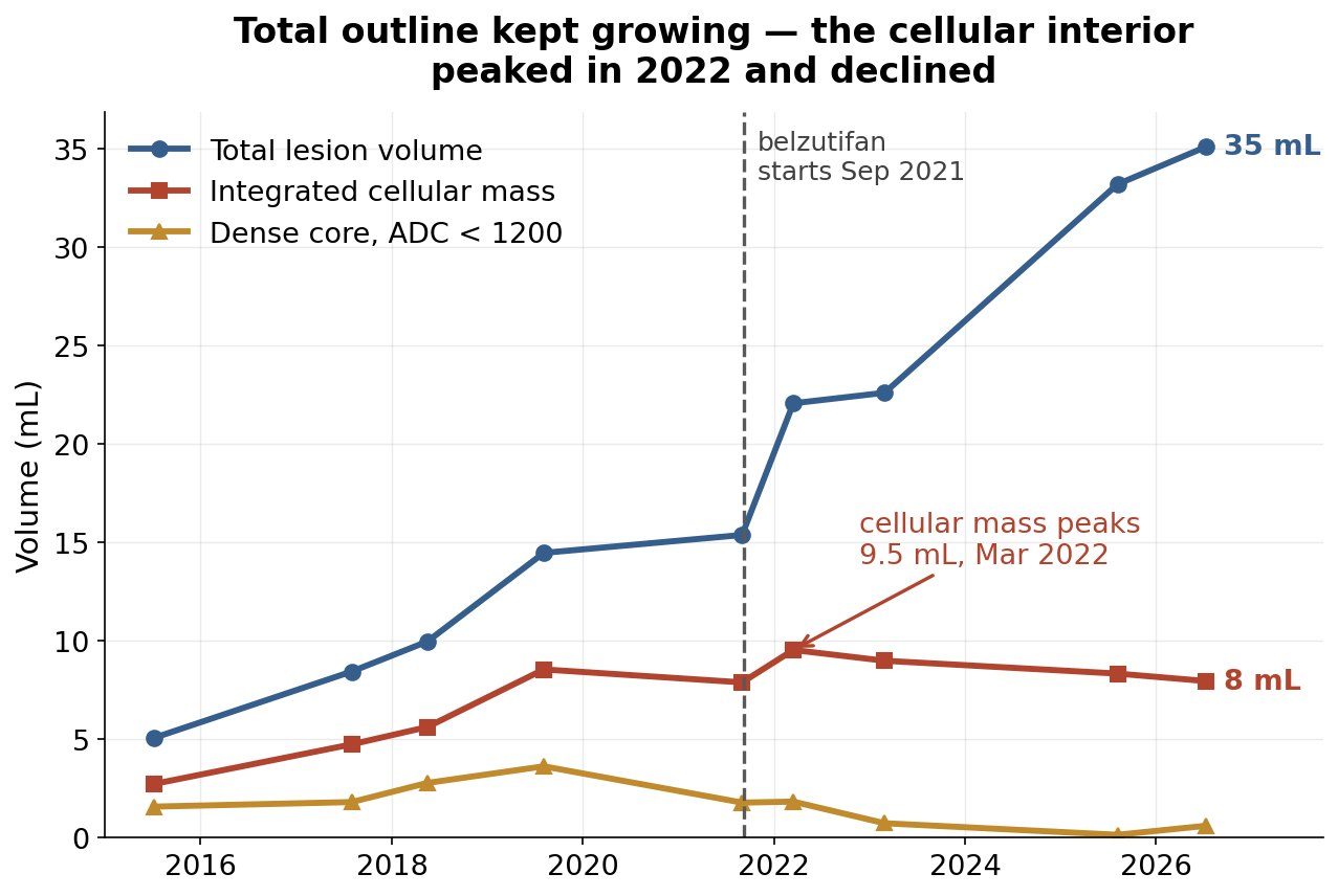
Before treatment (2015–2019), total volume, integrated cellular mass and dense cellular core tripled in lockstep — what genuine growth looks like. On treatment they split: the outline kept rising while every tissue-level measure turned down.
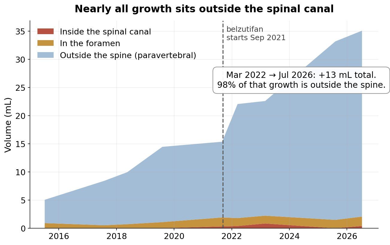
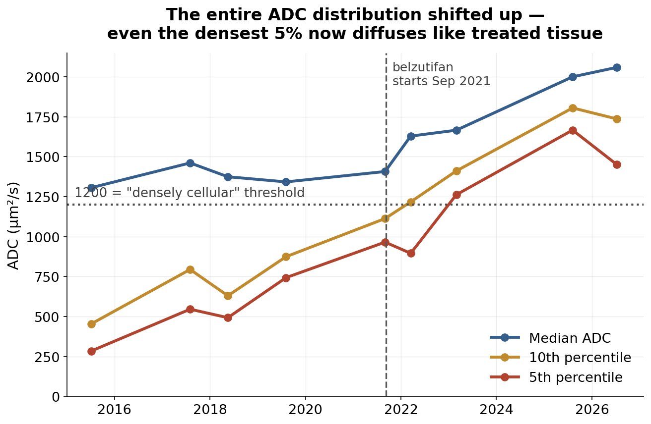
Integrated cellular mass peaked at 9.54 mL in March 2022 and declined to 7.96 mL by July 2026 (−17% on two-compartment diffusion mixing; −33% if the shrinking microvascular blood pool is subtracted first). The dense cellular core collapsed from its 2019 peak of 3.63 mL to 0.15–0.61 mL (−83% to −96%), reproducing the ~84% median solid-component regression of trial responders.[15] The dense core’s share of the total outline fell from 31% (2015) and 25% (2019) to 0.5–3.3% (2023–2026); every ADC percentile rose monotonically. Meanwhile 98% of the post-2022 volume gain accrued in the unconfined extradural (paravertebral) space; the component inside the spinal canal has been static for ten years (+0.02 mL), with ESCC 1c unchanged on every exam.
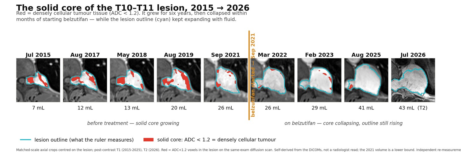
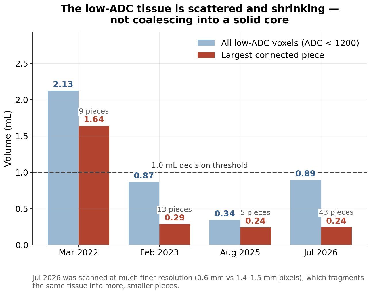
Three-dimensional connected-component analysis of dense voxels answers the resistant-clone question directly: the only contiguous ADC < 1.2 component ≥ 1 mL existed in March 2022 (1.64 mL) and collapsed to ~0.24 mL, flat since (2023: 0.29; 2025: 0.24; 2026: 0.24 mL largest). No contiguous dense focus ≥ 1 mL has existed since March 2022. A resistant subclone would appear here first — as one growing piece, not scattered rim flecks.
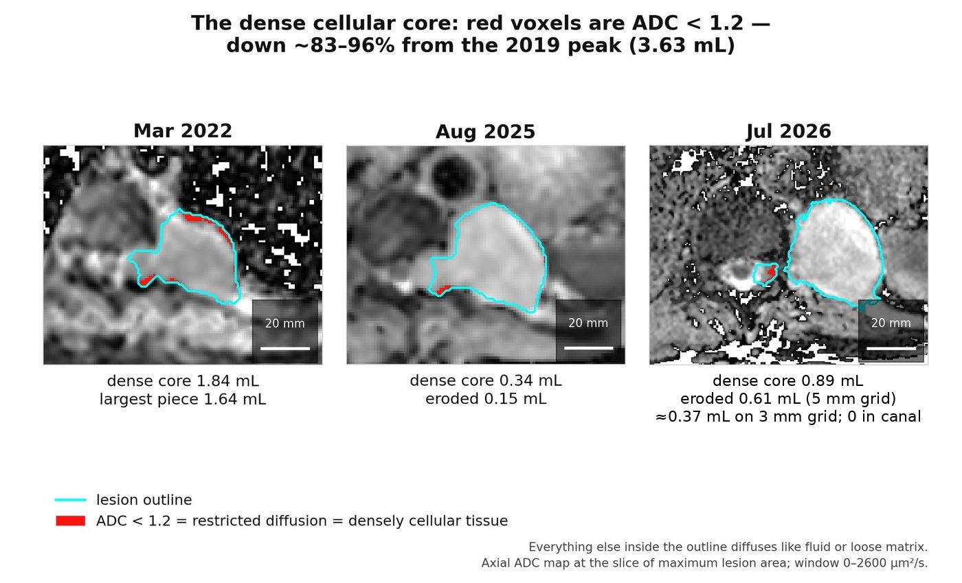
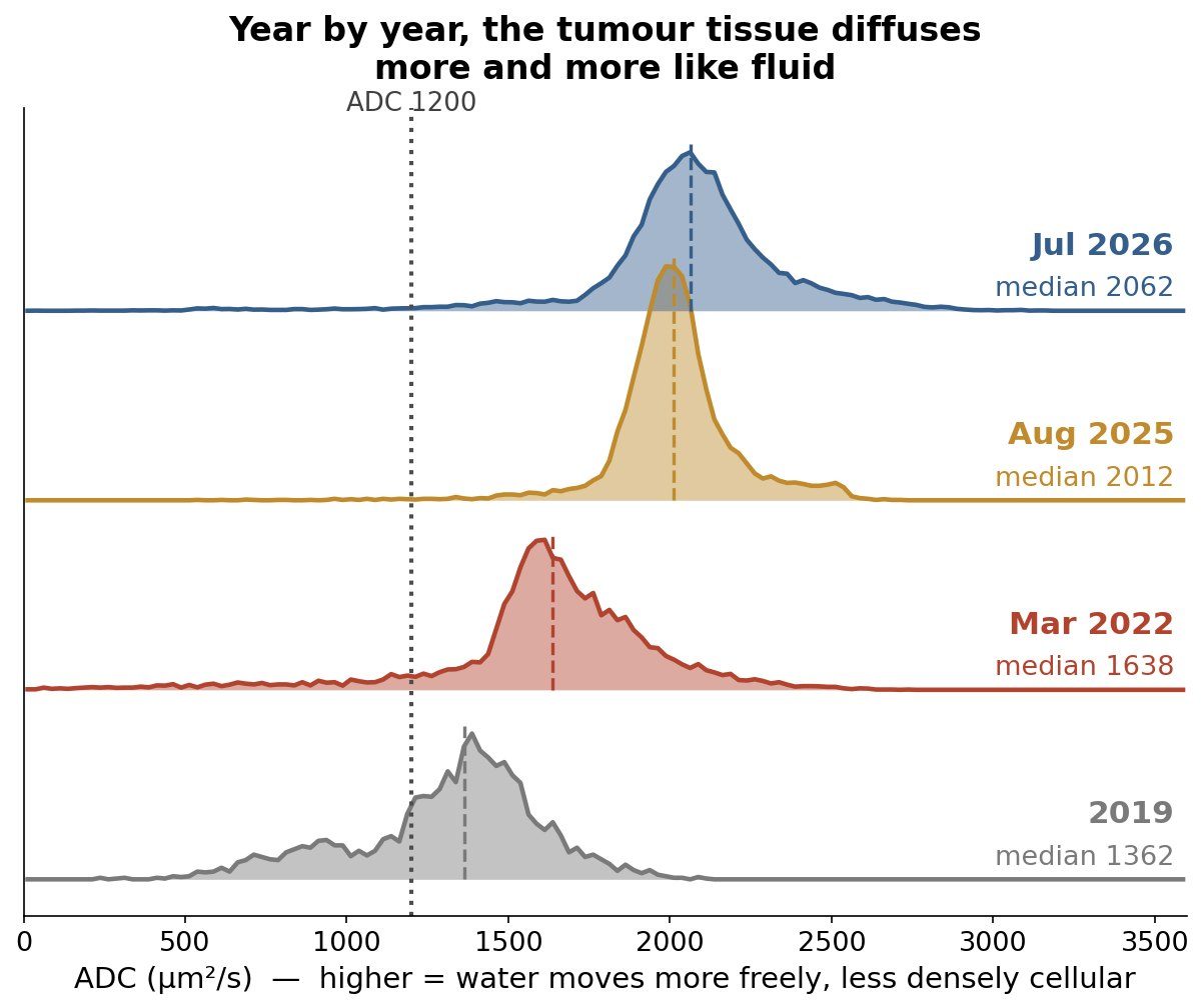
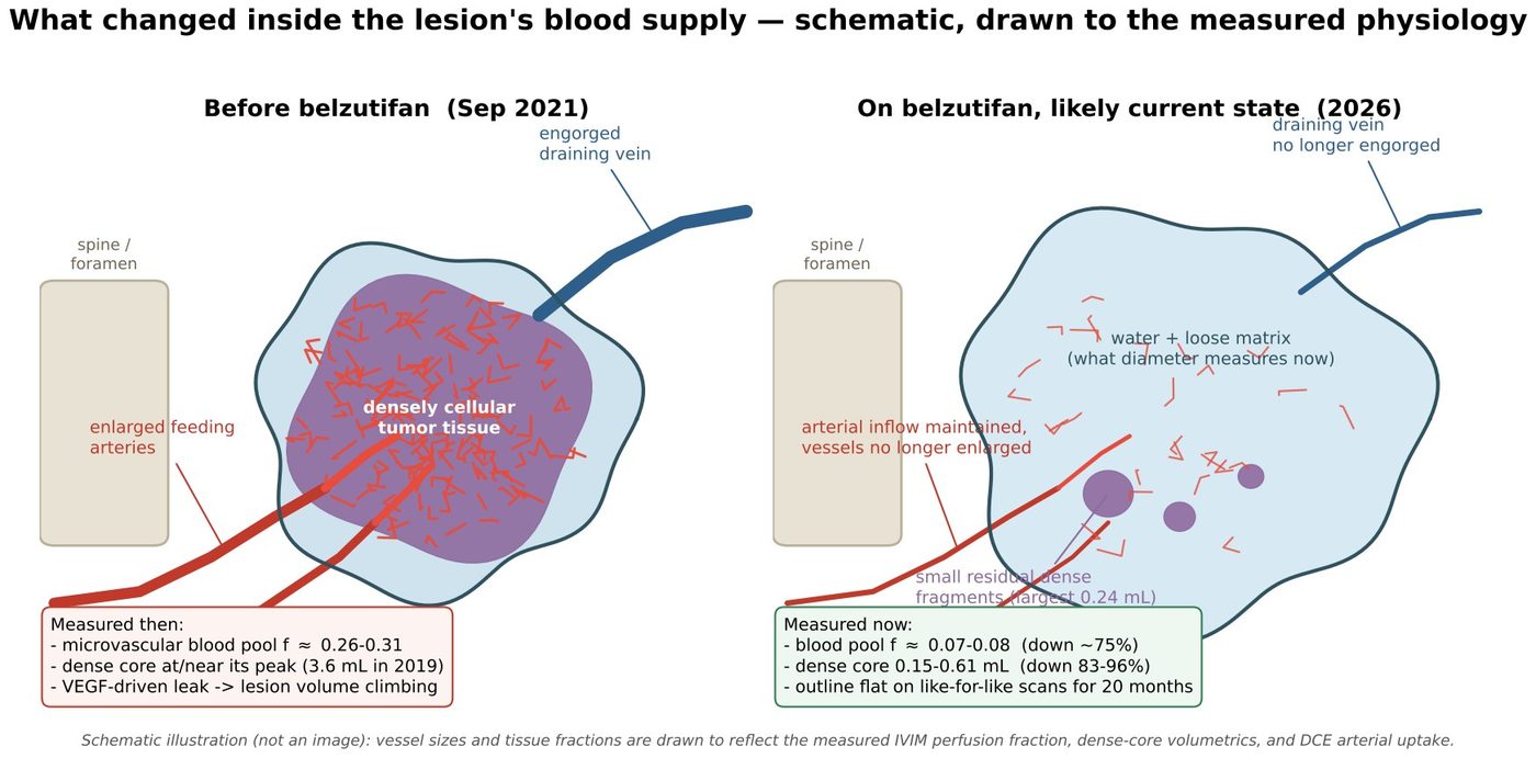
The ADC distribution of the whole lesion shifted toward free water: the median rose from 1362 (2019) to 2062 × 10−6 mm²/s (July 2026), and even the densest decile now sits above the dense-tissue cutoff. Within each comparable diffusion family the IVIM microvascular perfusion fraction collapsed (f ≈ 0.26–0.31 → 0.07–0.08), and contrast kinetics inverted from arterial flush-and-washout (2015–2019 arterial/venous/delayed relative enhancement ≈ 313/241/211%) to slow progressive accumulation (2025: 333/387/384%, delayed ≥ arterial). Intralesional T2 flow-void voxels fell to zero by 2024. The dominant feeder’s caliber persisted and arterial inflow did not fall — the lesion is not devascularized; rather, the immature VEGF-dependent exchange bed and its washout physiology have regressed, which is what HIF-2α/VEGF withdrawal should produce.[13,28,41] The radiology record carries independent corroboration: the March 2022 spine read noted the T5–T6 lesion’s “prominent feeding vessels… are less prominent.”
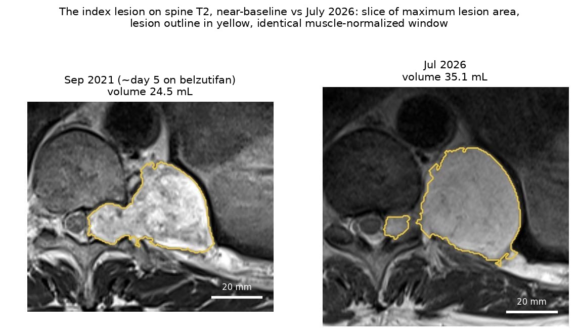
The decisive de-confounding comes from holding the protocol fixed. On the longitudinally consistent 5 mm axial T2 sequence, whole-lesion volume was 34.4 mL on 7 November 2024 and 35.1 mL on 10 July 2026 — a 20-month change of +0.7 mL (≈ +1%/yr; the extraspinal component −0.1%/yr). The widely quoted 43–44 mL figure for July 2026 comes from a same-day 3 mm high-resolution sequence that covers ~51 mm of craniocaudal extent versus ~31 mm routine and segments at a lower effective threshold. Wider coverage and the looser threshold each contribute to the same-day +9.0 mL difference: the ruler changed, not the lesion. A second convention — segmenting at half the lesion’s maximum signal — still gives ~6%/yr in the last interval. Read together, the honest statement is deceleration to at-or-near plateau on like-for-like measurement, a sharp break from the pre-treatment ~29%/yr.
| Date | Series family | Total outline (mL) | Cellular mass (mL) | Dense core ADC < 1.2 (mL) | ADC p10 |
|---|---|---|---|---|---|
| 2015-07 | abdominal 3-D T1 | 5.1 | 2.73 | 1.58 | 454 |
| 2017-08 | abdominal | 8.4 | 4.75 | 1.81 | 795 |
| 2018-05 | abdominal | 10.0 | 5.63 | 2.79 | 630 |
| 2019-08 | abdominal | 14.5 | 8.55 | 3.63 — peak | 874 |
| 2021-09-01 * | abdominal (truncated) | 15.4* | 7.89* | 1.79* | 1113 |
| 2021-09-14 | spine T2 (day ~5) | 24.5 | — | — | — |
| 2022-03 | abdominal / spine T2 | 22.1 / 20.2 | 9.54 — peak | 1.84 | 1218 |
| 2023-02 | abdominal / spine T2 | 22.6 / 26.8 | 8.99 | 0.74 | 1411 |
| 2023-08 | spine T2 | 33.4 | — | — | — |
| 2024-11 | spine T2 | 34.4 | — | — | — |
| 2025-08 | abdominal | 33.2 | 8.34 | 0.34 (0.15 eroded) | 1805 |
| 2026-07 | spine T2 (matched 5 mm) | 35.1 | 7.96 | 0.89 † (0.61 eroded) | 1737 (1804 eroded) |
| 2026-07 | spine T2 3 mm hi-res | 44.1 ‡ | — | ≈0.37 (0.8%; 0.00 canal/foramen) | 1861 |
Robustness: under loose, middle and tight masks alike, cellular mass peaks in March 2022 and declines (10.43→8.59 / 9.54→7.96 / 8.74→7.27 mL) and the dense core collapses 75–96% from the 2019 peak. Across a 16-combination grid of assumed diffusivities, 12 show declining cellular mass from the March 2022 baseline; only free-water-at-body-temperature ceilings produce growth. Cross-scanner harmonization against same-scan paraspinal muscle preserves the trend across four scanners and eleven years. The claim robust to every plausible assumption is no material cellular growth over five years on treatment; the decline is the likely, constants-dependent reading.
RECIST was built for cytotoxic chemotherapy in malignant tumors: response = shrinkage (≥30% in summed diameters), progression = ≥20% growth. Three properties of this setting strain those assumptions, and this case displays all three at once.
The earliest true responses are de-permeabilization phenomena — edema off, leak off, perfusion down — that no diameter captures.[41]
In the registration study’s own CNS data, cyst-inclusive reading gives a 44% objective response rate where solid-only reading of the same patients gives 76%.[15]
Size can rise while tumor regresses — this lesion’s exact behavior, inside a fibrous extradural boundary tethered to rib head, vertebral body and pleura that cannot collapse inward the way a millimeter-scale intradural nodule can.
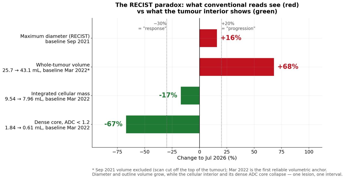
Read this way, the case stops being anomalous. A solid, cyst-free hemangioblastoma “progressing” on belzutifan is undocumented: a structured review of the indexed literature (PubMed, October 2026) found no published report of confirmed progression of the solid component of a CNS hemangioblastoma on belzutifan. Apparent enlargement in this disease is characteristically cyst-driven — which is why the registration study scored responses both cyst-inclusive and solid-only[15] — and the one real-world series that separates target from non-target lesions states that progression occurred only as “the emergence of new lesions,” with “no progression… in the primary target lesions.”[43] What the measurements suggest this lesion is doing — accumulating treatment-associated fluid while its tumor tissue regresses — resembles the familiar cyst phenomenon of CNS hemangioblastoma[9,10,15] expressed interstitially, inside an encapsulated extradural boundary, rather than as a loculated cyst. The fluid simply lacks a cyst wall to make the distinction obvious on a routine read; only tissue-level measures (diffusion, perfusion, enhancement kinetics) separate the compartments.
No radiology report or clinician in five years has ever recorded a sign of improvement in this lesion — not a measurement artifact so much as a gap in what the standard instruments can see — a change that diffusion, perfusion and compartment analysis do register.Patient-led analysis, October 2026
The March 2022 exam captures the inversion in miniature: a single scan that one neurosurgical consultation read as growth warranting resection, the institutional radiology report read as “essentially stable,” and the tissue-level measurements identify as the cellular compartment’s peak and turning point. Each read was correct on its own terms. That no reader ever saw improvement is the point, not a footnote: the standard instruments appear to have little sensitivity to this drug’s mechanism of action in solid tissue, which is reason to test whether size change should be the primary outcome at all.
The reporting standard now being proposed for this disease — report enhancing nodule, cyst and edema volumes separately rather than as one summed measurement — would be a substantial improvement. It is also, on the evidence of this lesion, incomplete in one specific way. Those are three volumes. A lesion that has lost its cellular fraction under HIF-2α inhibition and gained interstitial water without forming a cyst is scored entirely as nodule, reported as stable or slowly progressive on size, and the largest change in it does not enter the record at all. That is this case.
Median apparent diffusion coefficient within this lesion was flat at 1.37 and 1.36 (×10−3 mm2/s) in 2015 and 2019 — unchanged through a threefold increase in volume — and then rose to 1.42, 1.64, 1.68, 2.01 and 2.07 at September 2021, 2022, 2023, 2025 and 2026. Over the September 2021 to February 2023 interval, on the same scanner at the same b-values, the rise was 18% against a skeletal-muscle reference of −2%. The non-enhancing share fell over the same period, 2.8% to 0.6%: no cyst formed.
A rise in diffusivity inside a hypervascular tumor is ambiguous on its own, because blood is also mobile water, and conventional monoexponential diffusion imaging cannot separate a lesion filling with interstitial fluid from one filling with blood. Here intravoxel incoherent motion imaging separated them, and the two terms moved in opposite directions: tissue diffusivity rose from 1.0–1.2 to approximately 2.0 while the perfusion fraction collapsed from 0.26–0.31 to 0.07–0.08, which is skeletal-muscle level. Water up, blood down.
The suggestion this case supports is therefore a fourth reported quantity: within the enhancing volume, tissue diffusivity reported together with perfusion fraction, against a same-exam reference tissue. It is a qualifier on the nodule volume rather than a fourth segmentation — the compartment has no boundary to outline — and acquisition and fitting should follow the published multi-organ consensus recommendations so that results remain comparable between sites.[44] Two things make this cheaper than it sounds: diffusion sequences are already acquired in most spine and brain protocols, so for many archived studies the measurement is retrospective; and the known weakness of the technique, that protocol heterogeneity produces non-comparable parameters, now has a published remedy.
What it would buy is the distinction that volume cannot make. A lesion whose cellular fraction is falling while its volume rises may be a responding lesion filling with water. A lesion whose cellular fraction holds while its volume rises is more likely a tumor that is still growing. Those two situations call for opposite decisions, they are indistinguishable on diameter, and at present there is no prospective means of telling them apart.
Three limits should be stated with it. This is one lesion. It is extradural, where leaked plasma distributes through the tumor’s own interstitium because that space has lymphatic drainage and no containing envelope; an intradural lesion, where the same leak pools against a glial wall and forms a cyst, may partition its fluid differently, so the generalization to intramedullary or posterior fossa disease is untested. And the one published intratumoral study of a HIF-2α inhibitor, in metastatic renal carcinoma, reported that diffusion imaging was not informative[28] — with the monoexponential readout that is precisely the one expected to fail in a hypervascular lesion. Whether the separated parameters behave differently in a more vascular, more stromal tumor is untested in either direction. This is offered as a hypothesis with a worked example attached, not as a validated biomarker.
A patient on the only approved drug for this disease can be steered toward spinal surgery by a measurement convention. In this case the recommendation to resect came six months into treatment, on the exam that tissue-level analysis identifies as the turning point. Decisions anchored to total size alone risk operating on water.
The project’s primary outcome variable is the number of surgical procedures a person with VHL hemangioblastoma undergoes in a lifetime. Disability in this disease accrues across repeated CNS surgery,[11,12] and the strongest real-world signal for belzutifan is a fall in VHL-related procedures from 0.35 to 0.08 per patient-year, a reduction of roughly three-quarters.[18] If the instruments used to judge response cannot see response, that outcome is put at risk by the measurement itself — not by the disease or the drug.
That is why Direction 1 — three-compartment reporting, with nodule, cyst/fluid and edema measured and reported separately — and Direction 3 — a growth model that can certify a lesion safe to watch — sit first in the research agenda. Both are tractable with existing imaging; neither requires a new drug. The measurement lesson of the registration study (count the solid, not the fluid[15]) needs to be extended to encapsulated lesions that accumulate the same fluid interstitially, and mechanism-concordant markers — perfusion fraction, diffusion percentiles, enhancement kinetics, protocol-locked volumes, procedures per year, symptom burden — need to be tested as the decision variables for this drug class.
No management decisions have been made; nothing is scheduled or agreed. The central request is human, independent re-analysis, because the single largest limitation of the analysis is that it is one patient-side pipeline.
This is a patient-led synthesis compiled with large-language-model assistance and independent cross-review, with citations verified against PubMed; it is structured preparation for clinical judgment, not a substitute for it. All imaging analyses are self-derived with research-grade processing, not radiologist reads. Segmentations come from one analyst pipeline (three mask variants tested; no independent re-segmentation). The index-lesion record spans four scanners and multiple diffusion protocols; harmonization was checked but is imperfect. The September 2021 baseline is truncated. Cellular-mass estimates are model-dependent in absolute terms. Small-lesion measurements (1.4–6 mm on 3 mm slices) carry large relative uncertainty; pancreatic lesions are arterial-timing-dependent and their course after six months is genuinely indeterminate; the renal site carries prior ablation and surgical change. The index-lesion diagnosis is clinico-radiographic, without tissue (~85–95% confidence; an alternative differential of schwannoma was raised by the treating team in 2023). Boundary-erosion magnitudes depend on the measurement grid. The 2021 report of the patient’s earlier course is a non-peer-reviewed preprint.[39]
Citation metadata machine-verified against PubMed (NCBI), October 14, 2026; DOIs included for retrieval. Reference 39 is a preprint.